Corporate
 Top
Top
#08
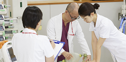
Improving Interventional Treatment Quality
Pursuing Intravascular Imaging Technology
Vital to coronary intervention:
About intravascular imaging
Among heart diseases, ischemic conditions including myocardial infarction and angina are said to be caused by lifestyle or aging, which results in arteriosclerosis (hardening) and stenosis (narrowing) of the coronary artery. Interventional therapy is a treatment in which a catheter is introduced through a radial (wrist) or femoral (thigh) blood vessel, to expand by balloon or stent the diameter of a coronary artery or a lower limb that has narrowed or become blocked; this has the effect of improving blood flow. Intravascular imaging (imaging diagnosis) is a technology that plays an important role in the interventional treatment of ischemic heart disease, by more clearly showing the disease state. The technology is used to provide high-definition images of the lumen (inside) of blood vessels, enabling physicians to understand the nature of blockages or arteriosclerosis. This way, they can diagnose the disease state and determine what interventional therapy to perform, and then evaluate stent placement after treatment.
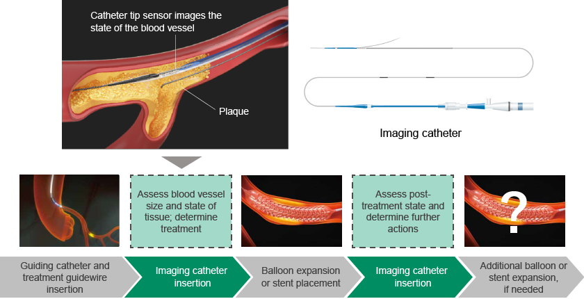
The role of intravascular imaging within interventional therapy
Intravascular imaging has been widely adopted in Japan; it is used in 90% of percutaneous coronary intervention (PCI) cases. It has become the norm for physicians to assess the state of lesions prior to treatment, making intravascular imaging indispensable to interventional therapy in Japan.
Two ways to obtain high-definition images:
Intravascular ultrasound (IVUS) and optical (OFDI)
There are two ways to perform intravascular imaging: Intravascular Ultrasound (IVUS), and Optical Frequency Domain Imaging (OFDI).
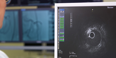
With IVUS, the reflections of ultrasound waves are used to provide cross-sectional images of the insides of blood vessels. To obtain the intravascular images, a narrow catheter containing a miniature sensor in its tip is inserted into the coronary artery and guided to the lesion (the place that is narrowed or blocked). The ultrasound waves produced there by the sensor are converted into images. IVUS enables deep observation by scanning widely from the blood vessel’s inner membrane through to its outer layers. Because the method does not require contrast agent to remove blood cells, the disease state can be observed in real time, regardless of the patient’s kidney function. This makes it the most common method of examination performed when placing a stent or expanding a balloon.
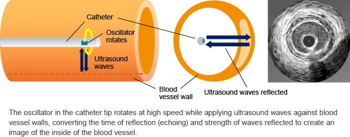
How an imaging catheter obtains images inside a blood vessel
OFDI uses near-infrared rays to take a high-resolution image of the inside of the blood vessel. Similarly to IVUS, a catheter containing a sensor in its tip is inserted into the blood vessel, where it emits near-infrared rays and uses what is called the "interference method"* to measure cross sections of the blood vessel and obtain images. OFDI can show differences in blood vessel wall tissues that cannot be detected with IVUS, enabling more detailed observation of the thickness of calcification or fibrous membrane tissue. Therefore, it is used primarily after treatment to evaluate things like stent placement.
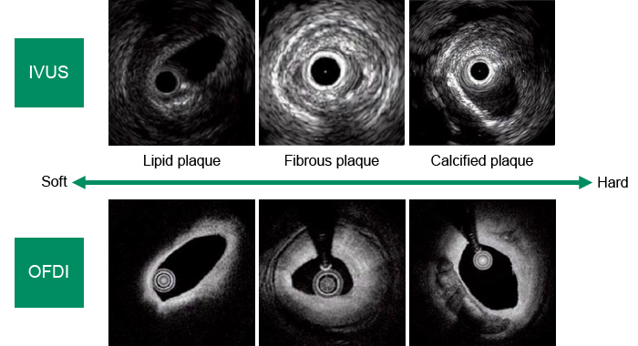
IVUS and OFDI image comparison
Terumo uses both of these technologies to provide Imaging Systems available nowhere else, which feature imaging instruments capable of producing high-definition images, and highly advanced catheters. Terumo is also continually enhancing its lineup of treatment devices to meet a variety of needs and contribute to more safe and effective interventional therapies, including the balloon catheters and stents required to broaden blood vessel width.
*
Interference method: Layering multiple lights to strengthen and weaken light waves
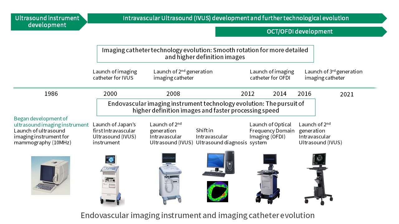
The First IVUS in Japan
Success by Believing in the Technology
Terumo Imaging Systems development began with the acquisition of ultrasound technology around 30 years ago. In the 1980s, the company launched its ultrasound diagnosis instrument for breast cancer screening. Normally, ultrasound instruments measured in the frequencies between 3.5 and 7Mhz. Terumo developed an instrument using the 10MHz frequency to enable diagnosis of breast cancer from the body surface. However, breast cancer screenings were not reimbursed by insurance at the time, leading to the company having no choice but to withdraw rom the market due to lack of growth. Later on, it began development of a miniature ultrasound probe for laparoscopic surgery that would use the same 10MHz ultrasound technology, but the project was ended due to lack of commercialization potential.
That was when Terumo looked to IVUS. At the time when the company was looking for ways to realize the potential of its ultrasound technology, the healthcare market had seen an increase in heart intervention, leading to increased need for IVUS in the United States and progress toward introducing it in Japan as well. Inside the company, research and development was progressing on advanced catheters and guidewires used to access and treat lesions during interventional procedures. That pushed Terumo to move forward in developing an IVUS that could use its ultrasound technology.
At the time, there was an issue with IVUS in which the images produced would become distorted at curved locations in blood vessels. Terumo set out to develop an imaging system that could enter any blood vessel and obtain a stable image, with a focus on not only high image quality but also ease of catheter entry and stability of image acquisition. The ultrasound imaging system that Terumo launched in 2000 was Japan’s first such product, and succeeded in achieving a dedicated catheter with the narrowest diameter and high durability, to go along with the imaging instrumentation. The technology cultivated through the IVUS development then led to development of OFDI technology in which the sensor could rotate at high speed without affecting catheter stability in obtaining an image.
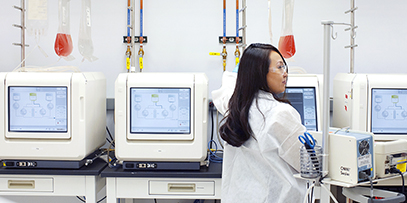
Terumo intravascular imaging technology continues to evolve today. Equipped with higher image definition and faster processing speed, the latest imaging systems enable shorter preparation, diagnosis, and imaging times, contributing to safer and more efficient interventional treatments. Terumo also meets the needs of physicians by developing specialized catheters that can be used in difficult cases and to access lower peripheral lesions from the wrist for treatment.
Creating New Value
For Interventional Therapy
IVUS and OFDI each have strengths and shortcomings, and there are limits to the information that just one of the two can obtain. Terumo is developing a dual sensor system (DSS) that combines both IVUS and OFDI technologies.
And as treatment and IT technologies evolve, new needs are emerging for imaging devices as well. Terumo is looking to the future in developing instruments that provide even greater added value, by utilizing AI to support IVUS imaging and suggest treatments and options to physicians as a treatment guide. Terumo engineers will continue to meet the challenge of providing technologies that meet the needs of medical professionals, for things like greater usability and medical cost efficiency, to contribute to better interventional therapy.
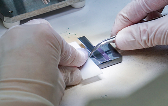
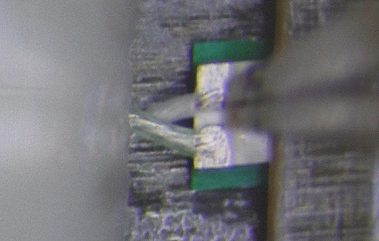
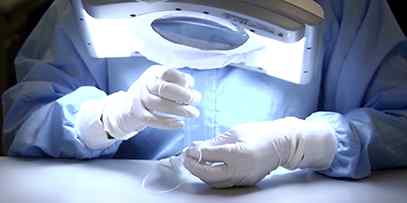
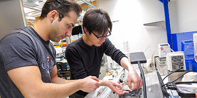
Welding an imaging catheter sensor under a microscope